Math Is Fun Forum
You are not logged in.
- Topics: Active | Unanswered
- Index
- » Science HQ
- » Pons
Pages: 1
#1 Yesterday 16:55:41
- Jai Ganesh
- Administrator

- Registered: 2005-06-28
- Posts: 52,716
Pons
Pons
Gist
The pons is a crucial part of the brainstem (linking the cerebrum to the cerebellum and spinal cord) that manages unconscious functions like breathing, sleep cycles, and relaying sensory/motor signals, containing nerve pathways and nuclei for facial sensation, hearing, balance, and other head/face functions, and is also the name of a popular dictionary/translation service.
The main function of the pons, a part of the brainstem, is to act as a relay station, connecting the cerebrum and cerebellum, and controlling vital functions like breathing, sleep cycles, facial expressions, and relaying sensory/motor signals for hearing, taste, balance, and movement coordination. It helps regulate arousal, fine motor control, and maintains equilibrium, essentially bridging communication between different parts of the brain and the body for essential processes.
Summary
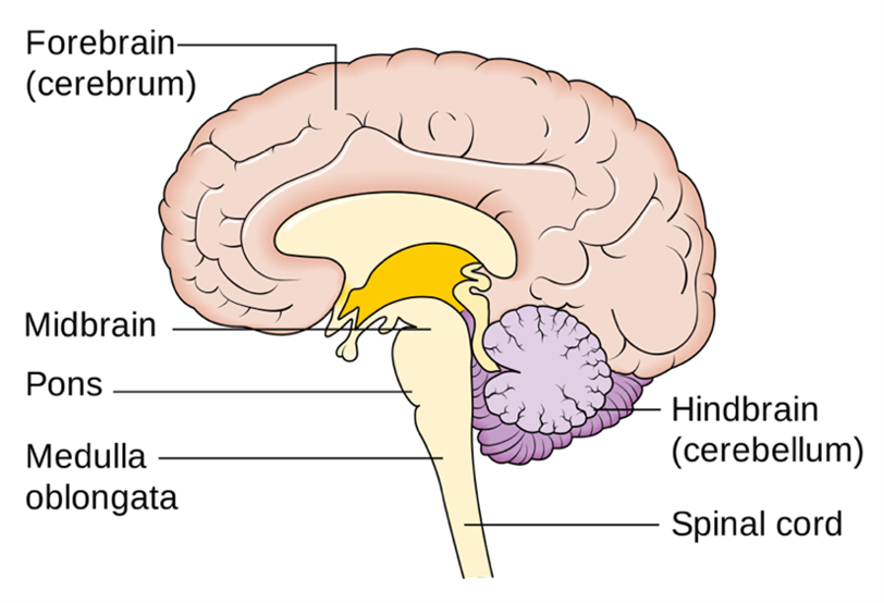
Pons is the portion of the brainstem lying above the medulla oblongata and below the cerebellum and the cavity of the fourth ventricle. The pons is a broad horseshoe-shaped mass of transverse nerve fibres that connect the medulla with the cerebellum. It is also the point of origin or termination for four of the cranial nerves that transfer sensory information and motor impulses to and from the facial region and the brain. The pons also serves as a pathway for nerve fibres connecting the cerebral cortex with the cerebellum.
The pons, while involved in the regulation of functions carried out by the cranial nerves it houses, works together with the medulla oblongata to serve an especially critical role in generating the respiratory rhythm of breathing. Active functioning of the pons may also be fundamental to rapid eye movement (REM) sleep.
Details
The pons (from Latin pons, 'bridge') is the part of the brainstem that, in humans and other mammals, lies inferior to the midbrain, superior to the medulla oblongata, and anterior to the cerebellum.
The pons is also called the pons Varolii ('bridge of Variolus'), after the Italian anatomist and surgeon Costanzo Varolio (1543–1575). The pons contains neural pathways and nerve tracts that conduct signals from the brain down to the cerebellum and medulla, as well as pathways that carry the sensory signals up into the thalamus.
Structure
The pons in humans measures about 2.5 centimetres (0.98 in) in length. It is the part of the brainstem situated between the midbrain and the medulla oblongata. The horizontal medullopontine sulcus demarcates the boundary between the pons and medulla oblongata on the ventral aspect of the brainstem, and the roots of cranial nerves 6, 7, and 8 emerge from the brainstem along this groove. The junction of pons, medulla oblongata, and cerebellum forms the cerebellopontine angle. The superior pontine sulcus separates the pons from the midbrain. Posteriorly, the pons curves on either side into a middle cerebellar peduncle.
A cross-section of the pons divides it into a ventral and a dorsal area. The ventral pons is known as the basilar part, and the dorsal pons is known as the pontine tegmentum.
The ventral aspect of the pons faces the clivus, with the pontine cistern intervening between the two structures. The ventral surface of the pons features a midline basilar sulcus along which the basilar artery may or may not course. There is a bulge to either side of the basilar sulcus, created by the pontine nuclei that are interweaved amid the descending fibres within the substance of the pons. The superior cerebellar artery winds around the upper margin of the pons.
Vasculature
Most of the pons is supplied by the pontine arteries, which arise from the basilar artery. A smaller portion of the pons is supplied by the anterior and posterior inferior cerebellar arteries.
Development
During embryonic development, the metencephalon develops from the rhombencephalon and gives rise to two structures: the pons and the cerebellum. The alar plate produces sensory neuroblasts, which will give rise to the solitary nucleus and its special visceral afferent (SVA) column; the cochlear and vestibular nuclei, which form the special somatic afferent (SSA) fibers of the vestibulocochlear nerve, the spinal and principal trigeminal nerve nuclei, which form the general somatic afferent column (GSA) of the trigeminal nerve, and the pontine nuclei, which relay to the cerebellum.
Basal plate neuroblasts give rise to the abducens nucleus, which forms the general somatic efferent fibers (GSE); the facial and motor trigeminal nuclei, which form the special visceral efferent (SVE) column; and the superior salivatory nucleus, which forms the general visceral efferent fibers (GVE) of the facial nerve.
Nuclei
A number of cranial nerve nuclei are present in the pons:
* mid-pons: the principal sensory nucleus of trigeminal nerve (5)
* mid-pons: the motor nucleus for the trigeminal nerve (5)
* lower down in the pons: abducens nucleus (6)
* lower down in the pons: facial nerve nucleus (7)
* lower down in the pons: vestibulocochlear nuclei (vestibular nuclei and cochlear nuclei) (8)
Function
Functions of these four cranial nerves (5–8) include regulation of respiration; control of involuntary actions; sensory roles in hearing, equilibrium, and taste; and in facial sensations such as touch and pain, as well as motor roles in eye movement, facial expressions, chewing, swallowing, and the secretion of saliva and tears.
The pons contains nuclei that relay signals from the forebrain to the cerebellum, along with nuclei that deal primarily with sleep, respiration, swallowing, bladder control, hearing, equilibrium, taste, eye movement, facial expressions, facial sensation, and posture.
Within the pons is the pneumotaxic center consisting of the subparabrachial and the medial parabrachial nuclei. This center regulates the transition from inhalation to exhalation.
The pons is implicated in sleep paralysis, and may also play a role in generating dreams.
Clinical significance
Central pontine myelinolysis is a demyelinating disease that causes difficulty with sense of balance, walking, sense of touch, swallowing and speaking. In a clinical setting, it is often associated with transplant or rapid correction of blood sodium. Undiagnosed, it can lead to death or locked-in syndrome.
Additional Information
Your pons is a part of your brainstem, a structure that links your brain to your spinal cord. It handles unconscious processes and jobs, such as your sleep-wake cycle and breathing. It also contains several junction points for nerves that control muscles and carry information from senses in your head and face.
Your pons is the second-lowest section of your brainstem, just above your medulla oblongata. It forms a key connection between your brain above it and your medulla oblongata and spinal cord below it.
Your pons is a key merging point for several of your cranial nerves, which are nerves with direct connections to your brain. Those nerve connections are vital, helping with several of the senses on or in your head, plus your ability to move various parts of your face and mouth.
Function:
What is the function of the pons?
Your pons is a part of your brainstem, which links your brain to your spinal cord. That makes your pons a vital section of your nervous system, providing a route for signals to travel to and from your brain. Several neurotransmitters in your pons facilitate brain function, particularly sleep.
Key jobs
Your pons handles several important jobs on its own.
* It influences your sleep cycle. Your pons sets your body’s level of alertness when you wake up.
* It manages pain signals. Your pons relays and regulates the signals that give you the sensation of pain from anywhere in your body below your neck.
* It works with other brain structures. Your pons is a key connection point to your cerebellum, another key part of your brain that handles balance and movement. It also works cooperatively with other parts of your brainstem that manage your breathing.
Cranial nerve connections
In addition, your pons contains several key junctions for four of your 12 cranial nerves, which are nerves that directly connect to your brain. Your cranial nerves (which use Roman numerals for their numbering) that connect to the pons are:
* Trigeminal nerve (Cranial Nerve V): Your trigeminal (try-gem-in-all) nerve provides the sense of touch and pain for your face and controls the muscles you use for chewing.
* Abducens nerve (CN VI): Your abducens (ab-DO-sens) nerve is one of the muscles that control eye movement. Damage to this nerve can cause double vision (diplopia).
* Facial nerve (CN VII): This nerve controls most of your facial expressions and your sense of taste from the front of your tongue.
* Vestibulocochlear nerve (CN VIII): Your vestibulocochlear (vest-ib-you-lo-co-klee-ar) nerve branches into your vestibular nerve and cochlear nerve. Your vestibular (vest-ib-you-lar) nerve gives you your sense of balance. Your cochlear (co-klee-ar) nerve gives you your sense of hearing.
How does it help with other organs?
Your pons helps with other organs by relaying sensory input and directly controlling some of your body’s unconscious processes. Those include your sleep-wake cycle and your breathing. Your ability to feel pain is also something your pons handles, and that sensation of pain can help you react to limit or prevent injuries.
Anatomy:
Where is the pons located?
Your pons is one of the lowermost structures in your brain, located near the bottom of your skull. It’s just above your medulla oblongata, which then connects to your spinal cord through the opening at the bottom of your skull.
What does it look like?
Your pons is a beige or off-white color. Its shape is much like the upper stem of a branch of cauliflower.
How big is it?
Pons’ dimensions are:
* Height: 1.06 inches tall (27 millimeters [mm]).
* Width: 1.49 inches (38 mm).
* Depth: 0.98 inches (25 mm).
What is it made of?
Like the rest of your brain and nervous system, your pons consists of various types of nervous system cells and structures. The nuclei (the plural term for “nucleus”) are nerves or clusters of brain cells that have the same job or connect to the same places.
Making up the nuclei are the following types of cells (with more about them below):
* Neurons: These cells make up your brain and nerves, transmitting and relaying signals. They can also convert signals into either chemical or electrical forms.
* Glial cells: These are support cells in your nervous system. While they don’t transmit or relay nervous system signals, they help the neurons that do.
Neurons
Neurons are the cells that send and relay signals through your nervous system, using both electrical and chemical signals. Each neuron consists of the following:
* Cell body: This is the main part of the cell.
* Axon: This is a long, arm-like part that extends outward from the cell body. At the end of the axon are several finger-like extensions where the electrical signal in the neuron becomes a chemical signal. These extensions, called synapses, lead to nearby nerve cells.
* Dendrites: These are small branch-like extensions (their name comes from a Latin word that means “tree-like”) on the cell body. Dendrites are the receiving point for chemical signals from the synapses of other nearby neurons.
* Myelin: This thin, fatty layer surrounds the axon of many neurons and acts as a protective covering.
Neuron connections are incredibly complex, and the dendrites on a single neuron may connect to thousands of other synapses. Some neurons are longer or shorter, depending on their location in your body and what they do.
Glial cells
Glial (pronounced glee-uhl) cells have many different purposes, helping develop and maintain neurons when you’re young and managing how the neurons work throughout your entire life. They also protect your nervous system from infections, control the chemical balance in your nervous system and create the myelin coating on the neurons’ axons. Your nervous system has 10 times more glial cells than neurons.
It appears to me that if one wants to make progress in mathematics, one should study the masters and not the pupils. - Niels Henrik Abel.
Nothing is better than reading and gaining more and more knowledge - Stephen William Hawking.
Offline
Pages: 1
- Index
- » Science HQ
- » Pons