Math Is Fun Forum
You are not logged in.
- Topics: Active | Unanswered
Pages: 1
#1 2026-01-22 18:44:20
- Jai Ganesh
- Administrator

- Registered: 2005-06-28
- Posts: 53,791
Dura Mater
Dura Mater
Gist
The tough outer layer of tissue that covers and protects the brain and spinal cord and is closest to the skull.
The meningeal layer of the dura mater creates several dural folds that divide the cranial cavity into freely communicating spaces. The function of the dural folds is to limit the rotational displacement of the brain. The folds include the following: The falx cerebri is a meningeal projection of dura in the brain.
Dura is the thick outer most layer of the 3 meninges. The thick fibrous dura surrounds, supports and protects the central nervous system (brain and spinal cord). In the cranium the dura forms folds to form partitions of the cranial cavity, and separates in places to form dural venous sinuses.
Summary
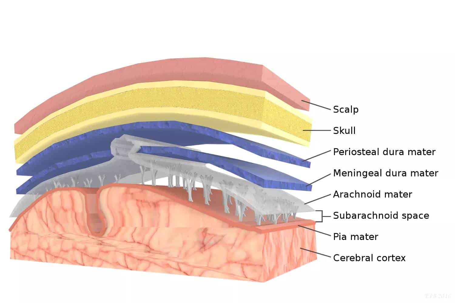
The dura mater (or just dura) is the outermost of the three meningeal membranes. The dura mater has two layers, an outer periosteal layer closely adhered to the neurocranium, and an inner meningeal layer known as the dural border cell layer. The two dural layers are for the most part fused together forming a thick fibrous tissue membrane that covers the brain and the vertebrae of the spinal column. But the layers are separated at the dural venous sinuses to allow blood to drain from the brain. The dura covers the arachnoid mater and the pia mater, the other two meninges, in protecting the central nervous system.
At major boundaries of brain regions such as the longitudinal fissure between the hemispheres, and the tentorium cerebelli between the posterior brain and the cerebellum the dura separates, folds and invaginates to make the divisions. These folds are known as dural folds, or reflections.
The dura mater is primarily derived from neural crest cells, with postnatal contributions from the paraxial mesoderm.
Details
The meninges are three layers of connective tissue that surround, support and protect the central nervous system (brain and spinal cord). From superficial to deep these layers are named the dura, arachnoid and pia. The dura mater is a thick, tough, fibrous membrane. It receives blood and nerve supply from the meningeal arteries, veins and nerves.
Cranial dura mater
In the cranium, the dura consists of two layers; an outer periosteal dura and an inner meningeal dura. The periosteal dura is closely attached to the internal surface of the skull bones while the inner meningeal dura is continuous with the dura of the spinal cord. The periosteal dura and meningeal dura are tightly fused together, except in a few places where they separate to form the dural venous sinuses; spaces that collect venous blood from the large veins of the brain. The dural venous sinuses are named as follows; superior sagittal sinus, inferior sagittal sinus, straight sinus, occipital sinus, transverse sinus, sigmoid sinus, marginal sinus, superior petrosal sinus, inferior petrosal sinus, petrosquamous sinus, cavernous sinus, sphenoparietal sinus, intercavernous sinus. The straight, occipital, transverse and superior sagittal sinuses all meet at the confluence of the sinuses. Arachnoid granulations, small tufts of arachnoid, protrude through the dura mater into the dural venous sinuses. They are the site of cerebrospinal fluid absorption into dural venous sinuses. Another feature of cranial dura are the dural folds. These are reflections of the inner meningeal dura which divide the cranium into separate compartments. The four dural folds are the falx cerebri, tentorium cerebelli, falx cerebelli, diaphragma sellae; in brief, they separate the cerebral and cerebellar hemispheres into divisions.
Spinal dura mater
In the spinal cord, only one layer of dura mater is found. Unlike in the cranium, the dura is not closely integrated with the overlying bones. Instead, a space exists between the dura and the vertebral bones known as the epidural space. The inferior aspect of the spinal dural sac is anchored to the coccyx by a thin connective tissue strand called the filum terminale.
Innervation
The innervation of the cranial dura mater is primarily sourced from the trigeminal (CN V) and vagus CN X) nerves, as well as spinal nerves C2/C3.
Trigeminal nerve
Several branches of the trigeminal nerve supply the dura mater:
* The tentorial branch of the ophthalmic nerve (CN V1), also known as the recurrent meningeal branch, supplies much of the supratentorial dura mater (i.e., posterior half of the falx cerebri, calvarial dura and superior surface of the tentorium cerebelli).
* The meningeal branch of the anterior ethmoidal nerve (branch of the nasociliary nerve (ophthalmic nerve)) supplies the central region of the anterior cranial fossa as well as anterior parts of the falx cerebri.
* The meningeal branch of the maxillary nerve (CN V2) provides innervation to the posterior region of the anterior cranial fossa as well as the anterior portion of the middle cranial fossa.
* The meningeal branch of the mandibular nerve (CN V3) supplies the posterolateral parts of the middle cranial fossa, before extending anteriorly to the lateral region of the anterior cranial fossa.
Vagus nerve
The posterior and lateral regions of the posterior cranial fossa and inferior surface of the tentorium cerebelli are all primarily innervated by the meningeal branch of the vagus nerve (which arises from its superior ganglion).
Spinal nerves C2/C3
The central region of the posterior cranial fossa, around the foramen magnum, receives innervation from sensory nerve fibers whose cell bodies are located in the spinal ganglia of spinal nerves C2/C3. The nerves may present as ascending branches of the meningeal branch of these spinal nerves which ascend the vertebral canal into the cranial cavity, via the foramen magnum. Alternatively, communicating branches of spinal nerves C2/C3 may pass to the hypoglossal nerve (CN XII), entering the cranial cavity via the meningeal branch of the hypoglossal nerve (via the hypoglossal canal). Some references occasionally also describe meningeal branches derived from the facial (CN VII) and glossopharyngeal (CN IX) nerves as supplying the posterior cranial fossa, however this remains debated.
Spinal dura
The innervation of the spinal dura mater is derived from a meningeal branch of each spinal nerve. It arises near the division of the spinal nerve into anterior and posterior rami, close to the gray/white rami communicating branches, before coursing through the intervertebral foramen into the vertebral canal.
Additional Information
The brain is an important part of the human body, as it performs vital functions and controls almost all bodily functions. It is protected by a strong framework of bones, called the skull. Besides this bony structure, it is also protected against several kinds of injuries or head traumas by the meninges, which are three-layered membranous structures that help protect the brain parts from damage.
The outermost layer of the meninges is the dura mater. The other two layers are collectively known as the leptomeninges and consist of the arachnoid mater (middle layer) and the pia mater (innermost layer). The dura mater is frequently known as the dura.
The dura protects its vital underlying components with a strong fibrous protective covering. In aspects of compressive impacts due to mass lesions and related edema, the compartmentalization of the brain through the help of dural reflections is especially important. The connection of the dural layers to each other, and the underlying leptomeninges and calvarium, has a huge impact on scientists’ knowledge of the impacts of intracranial injury.
Summary:
* The meninges safeguard the spinal cord and brain from tissue injury and assist the framework of the blood vessels
* Dura mater is on the outermost end of the meninges, situated directly beneath the skull and the bones of the vertebral column
* The dura mater is made up of two layers of connective tissue: the periosteal layer and the meningeal layer
* The periosteal layer lines the inner surface of the cranium’s bones. The periosteum is a dense fibrous membrane that covers the surface areas of bones
* The meningeal layer is found deep within the periosteal layer
* The dura mater has a vascular supply of its own
* The dura mater’s meningeal layer bends towards the inner direction of itself to form 4 structural forms, called the dural reflections
* The 4 dural reflections are: Diaphagma sellae, Falx cerebri, Tentorium cerebelli, and Falx cerebelli
* Dura mater encircles and continues to support the large venous channels (called dural sinuses) that help in carrying the blood from the brain to the heart.
* Haemorrhage, meningitis, and meningiomas are some complications involving the dura mater.
It appears to me that if one wants to make progress in mathematics, one should study the masters and not the pupils. - Niels Henrik Abel.
Nothing is better than reading and gaining more and more knowledge - Stephen William Hawking.
Offline
Pages: 1