Math Is Fun Forum
You are not logged in.
- Topics: Active | Unanswered
Pages: 1
#1 2024-10-04 22:12:43
- Jai Ganesh
- Administrator

- Registered: 2005-06-28
- Posts: 53,052
Cerebellum
Cerebellum
Gist
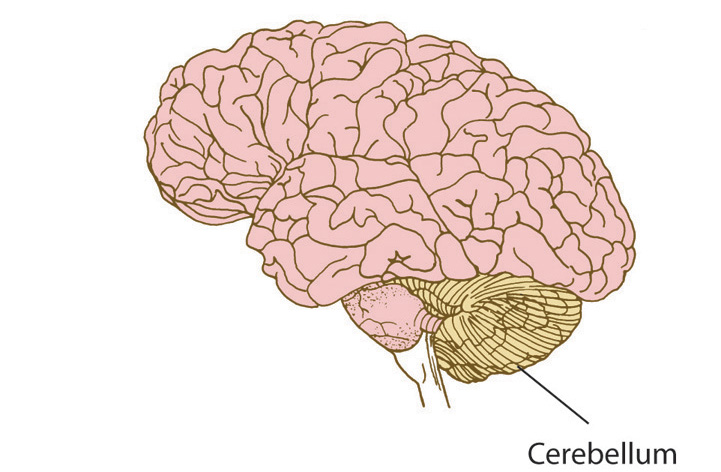
The cerebellum is a vital component in the human brain as it plays a role in motor movement regulation and balance control. The cerebellum coordinates gait and maintains posture, controls muscle tone and voluntary muscle activity but is unable to initiate muscle contraction.
It sits at the lower back of the brain, below, the rear cerebrum and behind the brain stem. It only accounts for around 10% of the brain's weight but contains up to 80% of all neurons in the organ. The cerebellum is primarily responsible for muscle control, including balance and movement.
Summary
The cerebellum (pl.: cerebella or cerebellums; Latin for "little brain") is a major feature of the hindbrain of all vertebrates. Although usually smaller than the cerebrum, in some animals such as the mormyrid fishes it may be as large as it or even larger. In humans, the cerebellum plays an important role in motor control and cognitive functions such as attention and language as well as emotional control such as regulating fear and pleasure responses, but its movement-related functions are the most solidly established. The human cerebellum does not initiate movement, but contributes to coordination, precision, and accurate timing: it receives input from sensory systems of the spinal cord and from other parts of the brain, and integrates these inputs to fine-tune motor activity. Cerebellar damage produces disorders in fine movement, equilibrium, posture, and motor learning in humans.
Anatomically, the human cerebellum has the appearance of a separate structure attached to the bottom of the brain, tucked underneath the cerebral hemispheres. Its cortical surface is covered with finely spaced parallel grooves, in striking contrast to the broad irregular convolutions of the cerebral cortex. These parallel grooves conceal the fact that the cerebellar cortex is actually a continuous thin layer of tissue tightly folded in the style of an accordion. Within this thin layer are several types of neurons with a highly regular arrangement, the most important being Purkinje cells and granule cells. This complex neural organization gives rise to a massive signal-processing capability, but almost all of the output from the cerebellar cortex passes through a set of small deep nuclei lying in the white matter interior of the cerebellum.
In addition to its direct role in motor control, the cerebellum is necessary for several types of motor learning, most notably learning to adjust to changes in sensorimotor relationships. Several theoretical models have been developed to explain sensorimotor calibration in terms of synaptic plasticity within the cerebellum. These models derive from those formulated by David Marr and James Albus, based on the observation that each cerebellar Purkinje cell receives two dramatically different types of input: one comprises thousands of weak inputs from the parallel fibers of the granule cells; the other is an extremely strong input from a single climbing fiber. The basic concept of the Marr–Albus theory is that the climbing fiber serves as a "teaching signal", which induces a long-lasting change in the strength of parallel fiber inputs. Observations of long-term depression in parallel fiber inputs have provided some support for theories of this type, but their validity remains controversial.
Details
The cerebellum, located in the lower back part of the brain, plays a vital role in most physical movements, including eye movements. Problems with the cerebellum can lead to coordination difficulties, fatigue, and other challenges.
This part of the brain helps a person drive, throw a ball, or walk across the room.
Problems with the cerebellum are rare and mostly involve movement and coordination difficulties.
The brain is a complex organ. It has three main parts; the cerebrum, the brainstem, and the cerebellum.
The cerebellum
The cerebellum is the lower-back part of the brain. It only accounts for around 10% of total brain weight but contains as many as 80% of all neurons in the brain.
The cerebrum
The cerebrum participates in higher levels of thinking and action. It is the largest part of the brain and covers the front, top, and upper back of the organ. Four lobes make up the cerebrum, each performing a different job.
* The frontal lobe: This sits at the front and top of the brain. It is responsible for the highest levels of human thinking and behavior, such as planning, judgment, decision making, impulse control, and attention.
* The parietal lobe: This lobe lies behind the frontal lobe. This lobe takes in sensory information and helps an individual understand their position in their environment.
* The temporal lobe: A lobe at the lower front of the brain. This lobe has strong links with visual memory, language, and emotion.
* The occipital lobe: This is at the back of the brain. The occipital lobe processes visual input from the eyes.
The brainstem
The brainstem is the bottom portion of the brain. It is below the cerebrum and connects to the spinal cord. The brainstem accompanies the cerebrum in promoting full physical and mental function.
The brainstem manages vital automatic functions, such as breathing, circulation, sleeping, digestion, and swallowing. These are the involuntary processes controlled by the autonomic nervous system. The brainstem also controls reflexes.
Cerebellum function
The cerebellum has several functions relating to movement and coordination, including:
* Maintaining balance: The cerebellum has special sensors that detect shifts in balance and movement. It sends signals for the body to adjust and move.
* Coordinating movement: Most body movements require the coordination of multiple muscle groups. The cerebellum times muscle actions so that the body can move smoothly.
* Vision: The cerebellum coordinates eye movements.
* Motor learning: The cerebellum helps the body to learn movements that require practice and fine-tuning. For example, the cerebellum plays a role in learning to ride a bicycle or play a musical instrument.
* Other functions: Researchers believe the cerebellum has some role in thinking, including processing language and mood. However, findings on these functions are yet to receive full exploration.
Disorders
As a result of the close relationship between the cerebellum and movement, the most common signs of cerebellar disorder involve a disturbance in muscle control.
Symptoms or signs include:
* lack of muscle control and coordination
* difficulties with walking and mobility
* slurred speech or difficulty speaking
* abnormal eye movements
* headaches
There are many disorders of the cerebellum, including:
* stroke
* brain bleeds
* toxins
* genetic anomalies
* infection
* cancer
Ataxia
The main symptom of cerebellum dysfunction is ataxia.
Ataxia is a loss of muscle coordination and control. An underlying problem with the cerebellum, such as a virus or brain tumor, can cause these symptoms. Loss of coordination is often the first sign of ataxia, and speech difficulties follow soon after.
Other symptoms include:
* blurry vision
* difficulty swallowing
* tiredness
* difficulties with precise muscle control
* changes in mood or thinking
Several factors can cause ataxia, including:
* genes
* poisons that damage the brain
* stroke
* tumors
* head injury
* multiple sclerosis
* cerebral palsy
* chicken pox and other viral infections
Sometimes ataxia is reversible when the underlying cause is treatable. In other cases, ataxia resolves without treatment.
Ataxia by toxins
The cerebellum is vulnerable to poisons, including alcohol and certain prescription medications.
These poisons damage nerve cells in the cerebellum, leading to ataxia.
The following toxins might cause ataxia:
* alcohol
* drugs, especially barbiturates and benzodiazepines
* heavy metals, including mercury and lead
* solvents, such as paint thinners
Alcohol consumption is the most common cause of toxin ataxia.
Ataxia disorders
Ataxia disorders are degenerative conditions. They can be either genetic or sporadic.
A genetic mutation causes genetic or hereditary ataxia. There are several different mutations and types.
These disorders are rare; even the most common type, Friedreich’s ataxia, affects only 1 in 40,000 people.
Sporadic ataxia is a group of degenerative movement disorders for which there is no evidence of inheritance. This condition usually progresses slowly and can develop into multiple system atrophy.
It presents a range of symptoms, including:
* fainting
* problems with heart rate
* erectile dysfunction
* loss of bladder control
These disorders usually get worse over time. There is no specific treatment to soothe or resolve symptoms, except in cases of ataxia where the cause is a vitamin-E deficiency.
There are several devices that can help people with irreversible ataxia, such as canes and specialized computers to support mobility, speech, and precise muscle control.
Additional Information
Cerebellum is section of the brain that coordinates sensory input with muscular responses, located just below and behind the cerebral hemispheres and above the medulla oblongata.
The cerebellum integrates nerve impulses from the labyrinths of the ear and from positional sensors in the muscles; cerebellar signals then determine the extent and timing of contraction of individual muscle fibres to make fine adjustments in maintaining balance and posture and to produce smooth, coordinated movements of large muscle masses in voluntary motions.
Like the cerebrum, the cerebellum is divided into two lateral hemispheres, which are connected by a medial part called the vermis. Each of the hemispheres consists of a central core of white matter and a surface cortex of gray matter and is divided into three lobes. The flocculonodular lobe, the first section of cerebellum to evolve, receives sensory input from the vestibules of the ear; the anterior lobe receives sensory input from the spinal cord; and the posterior lobe, the last to evolve, receives nerve impulses from the cerebrum. All of these nerve impulses are integrated within the cerebellar cortex. Three paired bundles of nerve fibres relay information to and from the cerebellum—the superior, middle, and inferior peduncles—which connect the cerebellum with the midbrain, pons, and medulla, respectively.
Functionally, the cerebellar cortex is divided into three layers: an outer synaptic layer (also called the molecular layer), an intermediate discharge layer (the Purkinje layer), and an inner receptive layer (the granular layer). Sensory input from different types of receptors is conveyed to specific regions of the receptive layer, which is made up of numerous small nerve cells that project axons into the synaptic layer. There the axons excite the dendrites of the Purkinje cells, which in turn project axons to portions of the four intrinsic nuclei (known as the dentate, globose, emboliform, and fastigial nuclei) and upon dorsal portions of the lateral vestibular nucleus. Most Purkinje cells use the neurotransmitter GABA and therefore exert strong inhibitory influences upon the cells that receive their terminals. As a result, all sensory input into the cerebellum results in inhibitory impulses’ being exerted upon the deep cerebellar nuclei and parts of the vestibular nucleus. Cells of all deep cerebellar nuclei, on the other hand, are excitatory (secreting the neurotransmitter glutamate) and project upon parts of the thalamus, red nucleus, vestibular nuclei, and reticular formation.
Injuries or disease affecting the cerebellum usually produce neuromuscular disturbances, in particular ataxia, or disruptions of coordinated limb movements. The loss of integrated muscular control may cause tremors and difficulty in standing.

It appears to me that if one wants to make progress in mathematics, one should study the masters and not the pupils. - Niels Henrik Abel.
Nothing is better than reading and gaining more and more knowledge - Stephen William Hawking.
Offline
Pages: 1