Math Is Fun Forum
You are not logged in.
- Topics: Active | Unanswered
Pages: 1
#1 2025-08-21 20:47:49
- Jai Ganesh
- Administrator

- Registered: 2005-06-28
- Posts: 53,831
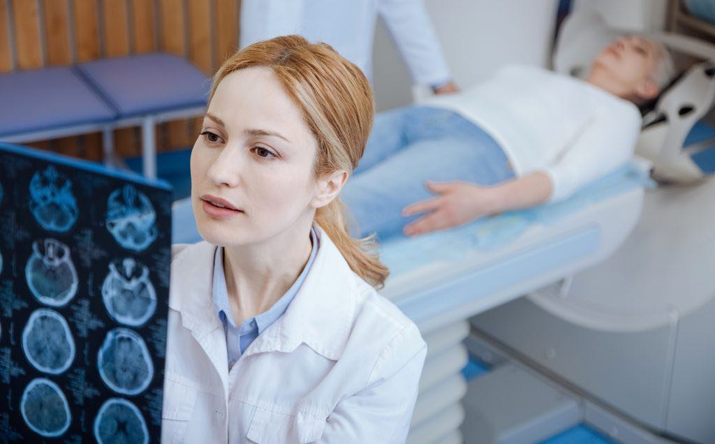
Computed Tomography
Computed Tomography
Gist
A CT scan (Computed Tomography) is an advanced X-ray imaging technique that uses a computer to create detailed, cross-sectional images of the body, allowing doctors to see internal structures like bones, blood vessels, and soft tissues. This non-invasive procedure produces more detailed images than traditional X-rays and helps diagnose, plan treatments for, and monitor various conditions, including tumors, injuries, and internal diseases.
A CT scan, or computed tomography scan, is a medical imaging technique used to create detailed cross-sectional images of the body. It's employed for a wide range of diagnostic and treatment-related purposes, including identifying injuries, tumors, infections, and other abnormalities.
Summary
Computed tomography (CT) is a diagnostic imaging method using a low-dose beam of X-rays that crosses the body in a single plane at many different angles.
CT was conceived by William Oldendorf and developed independently by Godfrey Newbold Hounsfield and Allan MacLeod Cormack, who shared a 1979 Nobel Prize for their inventions. A major advance in imaging technology, it became generally available in the early 1970s. The technique uses a tiny X-ray beam that traverses the body in an axial plane. Detectors record the strength of the exiting X-rays, and that information is then processed by computer to produce a detailed two-dimensional cross-sectional image of the body. A series of such images in parallel planes or around an axis can show the location of abnormalities and other space-occupying lesions (especially tumours and other masses) more precisely than can conventional X-ray images.
CT is the preferred examination for evaluating stroke, particularly subarachnoid hemorrhage, as well as abdominal tumours and abscesses.
Details
A computed tomography scan (CT scan), formerly called computed axial tomography scan (CAT scan), is a medical imaging technique used to obtain detailed internal images of the body. The personnel that perform CT scans are called radiographers or radiology technologists. CT scanners use a rotating X-ray tube and a row of detectors placed in a gantry to measure X-ray attenuations by different tissues inside the body. The multiple X-ray measurements taken from different angles are then processed on a computer using tomographic reconstruction algorithms to produce tomographic (cross-sectional) images (virtual "slices") of a body. CT scans can be used in patients with metallic implants or pacemakers, for whom magnetic resonance imaging (MRI) is contraindicated.
Since its development in the 1970s, CT scanning has proven to be a versatile imaging technique. While CT is most prominently used in medical diagnosis, it can also be used to form images of non-living objects. The 1979 Nobel Prize in Physiology or Medicine was awarded jointly to South African-American physicist Allan MacLeod Cormack and British electrical engineer Godfrey Hounsfield "for the development of computer-assisted tomography".
Types
On the basis of image acquisition and procedures, various type of scanners are available in the market.
Sequential CT
Sequential CT, also known as step-and-shoot CT, is a type of scanning method in which the CT table moves stepwise. The table increments to a particular location and then stops which is followed by the X-ray tube rotation and acquisition of a slice. The table then increments again, and another slice is taken. The table movement stops while taking slices. This results in an increased time of scanning.
Spiral CT
Spinning tube, commonly called spiral CT, or helical CT, is an imaging technique in which an entire X-ray tube is spun around the central axis of the area being scanned. These are the dominant type of scanners on the market because they have been manufactured longer and offer a lower cost of production and purchase. The main limitation of this type of CT is the bulk and inertia of the equipment (X-ray tube assembly and detector array on the opposite side of the circle) which limits the speed at which the equipment can spin. Some designs use two X-ray sources and detector arrays offset by an angle, as a technique to improve temporal resolution.
Electron beam tomography
Electron beam tomography (EBT) is a specific form of CT in which a large enough X-ray tube is constructed so that only the path of the electrons, travelling between the cathode and anode of the X-ray tube, are spun using deflection coils. This type had a major advantage since sweep speeds can be much faster, allowing for less blurry imaging of moving structures, such as the heart and arteries. Fewer scanners of this design have been produced when compared with spinning tube types, mainly due to the higher cost associated with building a much larger X-ray tube and detector array and limited anatomical coverage.
Dual energy CT
Dual energy CT, also known as spectral CT, is an advancement of computed Tomography in which two energies are used to create two sets of data. A dual energy CT may employ dual source, single source with dual detector layer, single source with energy switching methods to get two different sets of data.
* Dual source CT is an advanced scanner with a two X-ray tube detector system, unlike conventional single tube systems. These two detector systems are mounted on a single gantry at 90° in the same plane. Dual source CT scanners allow fast scanning with higher temporal resolution by acquiring a full CT slice in only half a rotation. Fast imaging reduces motion blurring at high heart rates and potentially allowing for shorter breath-hold time. This is particularly useful for ill patients having difficulty holding their breath or unable to take heart-rate lowering medication.
* Single source with energy switching is another mode of dual energy CT in which a single tube is operated at two different energies by switching the energies frequently.
CT perfusion imaging
CT perfusion imaging is a specific form of CT to assess flow through blood vessels whilst injecting a contrast agent. Blood flow, blood transit time, and organ blood volume, can all be calculated with reasonable sensitivity and specificity. This type of CT may be used on the heart, although sensitivity and specificity for detecting abnormalities are still lower than for other forms of CT. This may also be used on the brain, where CT perfusion imaging can often detect poor brain perfusion well before it is detected using a conventional spiral CT scan. This is better for stroke diagnosis than other CT types.
PET CT
Positron emission tomography–computed tomography is a hybrid CT modality which combines, in a single gantry, a positron emission tomography (PET) scanner and an X-ray computed tomography (CT) scanner, to acquire sequential images from both devices in the same session, which are combined into a single superposed (co-registered) image. Thus, functional imaging obtained by PET, which depicts the spatial distribution of metabolic or biochemical activity in the body can be more precisely aligned or correlated with anatomic imaging obtained by CT scanning.
PET-CT gives both anatomical and functional details of an organ under examination and is helpful in detecting different type of cancers.
Additional Information:
CT (Computed Tomography) Scan
A CT (computed tomography) scan is an imaging test that helps healthcare providers detect diseases and injuries. It uses a series of X-rays and a computer to create detailed images of your bones and soft tissues. A CT scan is painless and noninvasive. You might go to a hospital or imaging center for your CT scan.
Overview:
What is a CT (or CAT) scan?
A CT (computed tomography) or CAT (computed axial tomography) scan is a type of imaging test that helps detect diseases and injuries. It uses X-rays and a special computer to create detailed pictures of your bones, organs and soft tissues.
It creates images of the inside of your body by taking multiple X-ray pictures at different angles. When the images are combined, it creates a 3D image. This lets your provider see your body very clearly and in much more detail than a standard X-ray.
CT scans can show if a health condition is getting better or worse with treatment. It can detect things such as cancer and certain infections.
What does a CT scan show?
CT scans can show abnormalities like tumors, injuries and diseases in more detail than an X-ray. Examples include:
* Many types of cancer
* Pneumonia and emphysema
* Broken bones
* Heart disease
* Blood clots
* Bowel disorders (appendicitis, diverticulitis, blockages, Crohn’s disease)
* Kidney stones
* Brain injuries
* Spinal cord injuries
* Internal bleeding
Test Details:
How does a CT scan work?
Healthcare providers use CT scans to see things that regular X-rays can’t show. It produces detailed, clear and precise images of the organs and structures in your body. To get these images, a CT machine takes X-ray pictures as it moves around you.
X-rays alone take flat, 2D images. A CT scan takes several pictures at many angles to create cross-sectional images. Just like you can see the inside layers of a cake when you slice it, a CT can show the “layers” of your body. Taken together, the layers create a 3D image. Some CT scans use a contrast material (dye) to make the pictures even clearer.
Do I need to prepare for a scan?
Your healthcare provider will tell you everything you need to know before a CT scan. Here are some general guidelines:
* Plan to arrive early. Your provider will tell you when to come to your appointment.
* Wear comfortable clothes and remove any metal jewelry or clothing. Your provider may give you a hospital gown to wear.
Your provider might use contrast material to highlight certain areas of your body. Contrast helps make certain tissues, organs or blood vessels easier to see. For a CT scan with contrast, your provider typically injects the contrast (dye) into your vein through an IV. This dye can make you feel flushed or give you a metallic taste in your mouth. IV contrast agents usually flush from your system (when you pee) within 24 hours. Sometimes, you may be asked to drink a contrast liquid. This is especially helpful if you’re having a scan of your intestines.
Here are other things you may need to prepare for a CT scan with contrast:
* Blood test: You might need a blood test to make sure the contrast material is safe to use.
* Allergy medication: If you’re allergic to the contrast agent, you may need to take steroid and antihistamine medications the night before and the morning of your procedure. Be sure to check with your healthcare provider and have them order these medications for you if needed.
What should I expect during my CT scan?
During the test, you’ll usually lie on your back on a table (like a bed). When the scan begins:
* The bed moves into the doughnut-shaped machine. At this point, you’ll need to stay as still as possible because movement can blur the images.
* You may also be asked to hold your breath for a short period of time, usually fewer than 15 to 20 seconds.
* The scanner takes pictures of the area your healthcare provider needs to see. A CT scanner is relatively quiet.
* When the exam is over, the table moves back out of the scanner.
* A technologist trained specifically to perform CT scans will be there to guide you through the entire process.
How long does a CT scan take?
CT scans usually take about an hour. Most of that time is for the preparation. The scan itself usually takes fewer than five minutes.
Are there risks or side effects?
Healthcare providers consider CT scans safe. CT scans for children are safe, too. Like X-rays, CT scans use a small amount of ionizing radiation to capture images. The level of radiation you’re exposed to is small. If you have concerns about the health risks of CT scans, talk to your healthcare provider. They’ll help you make an informed decision about the scan.
CT scans themselves usually don’t cause side effects. But some people have side effects from the contrast material. These side effects may include:
* Nausea and vomiting
* Headaches
* Dizziness
Results and Follow-Up:
What happens after a CT scan?
You can go home after a CT scan. It’s safe to resume your normal activities. If your provider used contrast dye, they may tell you to drink lots of water.
The images from the CT scan are sent to a radiologist. They review your scans and prepare a report that explains the findings. Then, they send the report to the healthcare provider who ordered the CT.
When should I know my results?
It usually takes about 24 to 48 hours to get the results of your CT scan. In an emergency setting, like a hospital or emergency room, healthcare providers often receive results within an hour.
Once a radiologist and your healthcare provider have reviewed the results, you’ll either have another appointment or receive a call. Your healthcare provider will discuss the results with you.
What do the results of a CT scan mean?
It depends on what your healthcare provider was looking for. The report your radiologist writes may include things like:
* How they performed the CT (if contrast was used, positioning, what body parts they looked at)
* Any findings (descriptions of any irregularities or details as to what looked normal or abnormal)
* A summary called an Impression (including a possible diagnosis, the biggest finding, and recommendations for further study)
Sometimes, your provider will suggest additional testing after a CT. For example, they may recommend an MRI to get a different look at a suspicious area.
You may see words in the report that are unfamiliar to you. If you have questions about your CT report, don’t hesitate to ask your provider what it means. They’ll be able to explain how the findings affect your care.
It appears to me that if one wants to make progress in mathematics, one should study the masters and not the pupils. - Niels Henrik Abel.
Nothing is better than reading and gaining more and more knowledge - Stephen William Hawking.
Offline
Pages: 1